HOW(?) & WHY(?) Liquid-Eating & Intermittent-Fasting can be so beneficial to your Health...

Thursday, 10 July 2008

TSG = Transient Supernormal Glycemia ... Why ZERO symptoms following 100% blockage occlusion of coronary artery [0:32 to 0:55 mins] ...HOW ?


"Every Day And In Every Way I Am Getting Better And Better"...
.
.
.
Ann Med. 2008 May 16:1-7. [Epub ahead of print]Click here to read

Acute HYPERglycemia [TRANSIENT SUPERNORMAL GLYCEMIA (TSG)] induces a HEALTHY IMMUNE response in young HUMANS withOUT ... 'GM-INSULIN-TREATED diabetes' ...

Folkhalsan Institute of Genetics, Folkhalsan Research Center, Biomedicum Helsinki, Finland.

Background. Patients with GM INSULIN TREATED [GIT] diabetes are at a substantially increased risk of cardiovascular disease FOR SOME SPECIFIC REASON ...


Stress-induced HYPERglycemia, in turn, ALLEGEDLY APPEARS TO SHOW AN ALLEGED worsenING OF the prognosis of Patients suffering from an acute myocardial infarction [AKA FUEL-ANEMIA STARVATION OF HEART MUSCLE] ?

However, the mechanisms behind these APPARENT findings are ALLEGEDLY incompletely known.


Aim. To investigate whether markers of chronic [MONOTONOUS] inflammation, and oxidative stress respond to acute [SHORT TERM] HYPERglycemia in patients with GM INSULIN TREATED [GIT] diabetes.


Methods. The plasma glucose concentration was rapidly raised from 5 mmol/L [90MG/DL] to 15 mmol/L [270MG/DL] in 35 Males ...


22 Men with GM INSULIN TREATED [GIT] diabetes ...

and

13 age-matched non-Diabetic Volunteers ... and maintained AS TRANSIENT SUPERNORMAL GLYCEMIA [TSG] for 2 HOURS.


All Participants were young non-Smokers without any signs of diabetic or other complications.

Markers of chronic [MONOTONOUS] inflammation, and oxidative stress were analysed in serum/plasma samples drawn at base-line and after 120 min of HYPERglycemia.


Results. Compared to NORMOglycemia, acute HYPERglycemia increased the interleukin (IL)-6 concentrations by 39% in Patients with GM INSULIN TREATED [GIT] diabetes (P<0.01)


During HYPERglycemia the PROFOUNDLY BENEFICIAL superoxide dismutase [SOD] concentration was increased by ...
17% in the HUMAN Volunteers (P<0.01)
5% in the Patients with GM INSULIN TREATED [GIT] diabetes (P=NS)

The increase in tumour necrosis factor (TNF)-agr was larger in Patients with GM INSULIN TREATED [GIT] diabetes than in non-Diabetic volunteers (35% versus -10%, P<0.05).


Conclusions... This study shows that acute HYPERglycemia induces a HEALTHY IMMUNE response in HEALTHY HUMANS FREE FROM A GM INSULIN TREATMENT [GIT] ... THAT IS ALLEGEDLY FOR DiabetIC HEALTH PURPOSES.

Keywords: Acute HYPERglycemia; cardiovascular disease; inflammation; macrovascular disease; oxidative stress; GM INSULIN TREATMENT [GIT] diabetes
http://www.InformaWorld.cOM/smpp/content~db=all~content=a793151240

History of wound care AND HYPERGLYCEMIA LIKE HEALING VIA HONEY [AND SUGAR WORKS JUST AS WELL] ...

The history of wound care spans from prehistory to modern medicine. As wounds naturally heal by themselves, regardless of whether recovery from the scar or recovery from lost body tissue was a possibility, hunter-gatherers would have noticed several factors and certain herbal remedies would speed up or assist the process, especially if it was grievous. In ancient history, this was followed by the realisation of the necessity of hygiene and the halting of bleeding, where wound dressing techniques and surgery developed. Eventually the germ theory of disease also assisted in improving wound care.


Ancient medical practice

The treatment of acute and chronic wounds is an ancient area of specialization in medical practice, with a long and eventful clinical history that traces its origins to ancient Egypt and Greece. The Papyrus of Ebers, circa 1500 BC, details the use of lint, animal grease, and honey as topical treatments for wounds. The lint provided a fibrous base that promoted wound site closure, the animal grease provided a barrier to environmental pathogens, and the honey served as an antibiotic agent. The Egyptians believed that closing a wound preserved the soul and prevented the exposure of the spirit to "infernal beings," as was noted in the Berlin papyrus. The Greeks, who had a similar perspective on the importance of wound closure, were the first to differentiate between acute and chronic wounds, calling them "fresh" and "non-healing", respectively. Galen of Pergamum, a Greek surgeon who served Roman gladiators circa 120-201 A.D., made many contributions to the field of wound care. The most important was the acknowledgment of the importance of maintaining wound-site moisture to ensure successful closure of the wound. There were limited advances that continued throughout the Middle Ages and the Renaissance, but the most profound advances, both technological and clinical, came with the development of microbiology and cellular pathology in the 19th century.

Honey [AKA CONCENTRATED HYDRATED LIQUID SUGAR] ...

Honey’s antibacterial properties help promote healing infected wounds.[1] Honey used as an topical ointment.

19th century

The first advances in wound care in this era began with the work of Ignaz Philipp Semmelweis, a Hungarian obstetrician who developed sterile surgical procedures, and Louis Pasteur, a French scientist known as the "father of microbiology" for his germ theory of disease. Semmelweis's work was furthered by an English surgeon, Joseph Lister, who began treating his surgical gauze with carbolic acid, known today as phenol, and subsequently dropped his surgical team's mortality rate by 45%. Building on the success of Lister's pretreated surgical gauze, Robert Wood Johnson, co-founder of Johnson & Johnson, began producing gauze and wound dressings treated with iodine. These innovations in wound-site dressings marked the first major steps forward in the field since the advances of the Egyptians and Greeks centuries earlier. The next advances would arise from the development of polymer synthetics for wound dressings and the "rediscovery" of moist wound-site care protocols in the mid 20th century.

Wound-site dressing

1950s onward

With advancements in material and tissue sciences, the field of wound-site dressings increased considerably. The ability to bolster wound-site re-epithelialization has been improved as well as improving their clinical efficacy. With the advent of fibrous synthetics of nylon, polyethylene, polypropylene, and polyvinyls in the 1950s, researchers and doctors in the field of wound care are able to significantly accelerate the natural wound healing process. Following the research and articles of George Winter and Howard Maibach in the 1960s, testing the efficacy of synthetic "wet" polymer wound dressings, the 1970s and 1980s represented the dawn of modern wound care treatment. In the 1990s, improvements in composite and hybrid polymers expanded the number of available materials for wound dressings. These improvements, coupled with the developments in tissue engineering, have given rise to a new class of wound dressings called "living skin equivalents." Often cited as a misnomer because they lack key components of whole living skin, "living skin equivalents" represent the future of wound dressings and possess the potential to serve as cellular platforms for the release of growth factors essential for proper wound healing. Other new developments have been focusing on handling patients concerns. Affected individuals often report pain as dominant in their lives[2] and the pain associated with chronic wounds should be handled as one of the main management priorities. Symptomatic treatment of pain as part of patient centered concerns must go hand-in-hand with treating the underlying etiology or cause of the wound.

References

  1. ^ Peter Charles Molan (2001). "Honey as a topical antibacterial agent for treatment of infected wounds". Nurs Times 49 (7-8): 96.
  2. ^ Krasner, D. 1998. Painful venous ulcers: themes and stories about living with the pain and suffering. Journal of Wound, Ostomy, and Continence Nursing, Volume 25, Issue 3, Pages 158-168. Accessed January 1, 2007.

Sources

  • Ovington, L. G. (2002). "The evolution of wound management: ancient origins and advances of the past 20 years." Home Healthcare Nurse. 20, p 652-656.

  • Sipos, P., Gyory, H., Hagymasi, K., Ondrejka, P., and Blazovics, A. (2004). "Special wound healing methods used in ancient Egypt and the mythological background." World Journal of Surgery. 28, 211-216

External links


Long-chain fatty acids (LCFA) are the major
oxidation fuel of the healthy heart in vivo and in vitro,
while carbohydrates, especially lactate and glucose,
provide the remaining energy source (Neely et al. 1969,
Simonsen and Kjekshus 1978, Stanley et al. 1997).
However, heart metabolism can rapidly adapt to changes
in fuel-type availability. For example, the heart shifts to
preferential utilization of glucose when the arterial
concentration of free fatty acids falls below 0.3 mmol/l
(Nuutila et al. 1994).
A few studies analyzed the role of fuel-type
utilized by vascular tissues. It has been reported that
endothelial cells can use both glucose and fatty acids as
fuel-type substrate, and that they utilize triglycerides as
alternative fuel during glucose deprivation aka Relative-HYPOglycemai-distress (Culic et al.1999).

Wednesday, 9 July 2008

SOD = SuperOxide Dismutase


"Every Day And In Every Way I Am Getting Better And Better"...
.
.
.
Am J Hypertens. 2008 Jun;21(6):708-14. Epub 2008 Apr 10.Click here to read Links

Hepatic effects of a fructose diet in the stroke-prone spontaneously HYPERtensive rat.

Center for Metabolic Disease, Ordway Research Institute, Albany, New York, USA.

jbrosnan@ordwayresearch.org


BACKGROUND: Feeding stroke-prone spontaneously hypertensive rats (SHRSP) a diet rich in fructose results in a profound glucose intolerance not observed in the normotensive Wistar Kyoto (WKY) strain.

The aim of this study was to investigate the role of the liver in the underlying mechanisms in the SHRSP.

METHODS: SHRSP and WKY rats were fed either 60% fructose or regular chow for 2 weeks with blood pressure being measured using tail-cuff plethysmography and radiotelemetry.

Intraperitoneal glucose tolerance tests were performed and livers harvested for analysis of expression of inflammatory mediators and antioxidant proteins by western blotting and quantitative reverse transcriptase-PCR.

The serum triglyceride content and fatty acid profiles were also measured.

RESULTS: Feeding SHRSP and WKY on 60% fructose for 2 weeks resulted in glucose intolerance with no increases in levels of blood pressure.

Serum triglycerides were increased in both strains of fructose-fed rats with the highest levels being observed in the SHRSP.

The serum fatty acid profiles were changed with large increases in the amounts of oleic acid (18.1) and reductions in linoleic acid (18.2).

Levels of expression of c-jun N-terminal kinase/stress activated protein kinase (JNK/SAPK), and nuclear factor kappaB (NF-kappaB) were shown to be unchanged between the livers of the chow and fructose-fed groups.

In contrast, protein levels of the three isoforms of superoxide dismutase (SOD) were upregulated in liver of SHRSP fed on fructose while only manganese SOD (MnSOD) was upregulated in fructose-fed WKY rats.

CONCLUSIONS: These results demonstrate that the major contribution of the liver in the early pathogenesis of metabolic syndrome may be an increased secretion of triglyceride containing altered proportions of fatty acid pools.

Feeding rats a diet rich in fructose does not affect hepatic expression of inflammatory pathways and the increased hepatic SOD expression may constitute an early protective mechanism.

Feeding stroke-prone spontaneously hypertensive rats (SHRSP) a diet rich in fructose results in a profound glucose intolerance associated with increased hepatic SOD expression that is apparently profoundly protective.


SuperOxide Dismutase


Structure of the monomeric unit of human superoxide dismutase 2
Identifiers
Symbol SOD1
Alt. Symbols ALS, ALS1
Entrez 6647
HUGO 11179
OMIM 147450
RefSeq NM_000454
UniProt P00441
Other data
EC number 1.15.1.1
Locus Chr. 21 q22.1
Identifiers
Symbol SOD2
Entrez 6648
HUGO 11180
OMIM 147460
RefSeq NM_000636
UniProt P04179
Other data
EC number 1.15.1.1
Locus Chr. 6 q25
superoxide dismutase 3, extracellular
Identifiers
Symbol SOD3
Entrez 6649
HUGO 11181
OMIM 185490
RefSeq NM_003102
UniProt P08294
Other data
EC number 1.15.1.1
Locus Chr. 4 pter-q21

The enzyme SuperOxide Dismutase (SOD, EC 1.15.1.1), catalyzes the dismutation of superoxide into oxygen and hydrogen peroxide. As such, it is an important antioxidant defense in nearly all cells exposed to oxygen. One of the exceedingly rare exceptions is Lactobacillus plantarum and related lactobacilli, which use a different mechanism.

Contents

Reaction

The SOD-catalysed dismutation of superoxide may be written with the following half-reactions :

  • M(n+1)+ − SOD + O2 → Mn+ − SOD + O2
  • Mn+ − SOD + O2 + 2H+ → M(n+1)+ − SOD + H2O2.

where M = Cu (n=1) ; Mn (n=2) ; Fe (n=2) ; Ni (n=2).

In this reaction the oxidation state of the metal cation oscillates between n and n+1.

Types

General

SOD was discovered by Irwin Fridovich and Joe McCord, which prior were known as several metalloproteins with unknown function (for example, CuZnSOD was known as erythrocuprein). Several common forms of SOD exist: they are proteins cofactored with copper and zinc, or manganese, iron, or nickel.

Brewer (1967) identifed superoxide dismutase as an indophenol oxidase by protein analysis of starch gels using the phenazine-tetrazolium technique. Brewer detected this enzyme in several human tissues an indophenol oxidase A(IPO-A). After that Brewer observed an electrophoretic variant of IPO-A, which he called 'Morenci,' in 3 generations of a family with presumed male-to-male transmission. Using a RT-PCR analysis Brewer has identified 5 splice variants of SOD1. The variants were expressed in brain, a region involved in amyotrophic lateral sclerosis.

  • The cytosols of virtually all eukaryotic cells contain an SOD enzyme with copper and zinc (Cu-Zn-SOD). (For example, Cu-Zn-SOD available commercially is normally purified from the bovine erythrocytes: PDB 1SXA, EC 1.15.1.1). The Cu-Zn enzyme is a homodimer of molecular weight 32,500. The two subunits are joined primarily by hydrophobic and electrostatic interactions. The ligands of copper and zinc are histidine side chains.

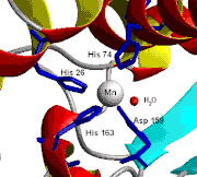
  • Chicken liver (and nearly all other) mitochondria, and many bacteria (such as E. coli) contain a form with manganese (Mn-SOD). (For example, the Mn-SOD found in a human mitochondrion: PDB 1N0J, EC 1.15.1.1). The ligands of the manganese ions are 3 histidine side chains, an aspartate side chain and a water molecule or hydroxy ligand depending on the Mn oxidation state (respectively II and III).

  • E. coli and many other bacteria also contain a form of the enzyme with iron (Fe-SOD); some bacteria contain Fe-SOD, others Mn-SOD, and some contain both. (For the E. coli Fe-SOD: PDB 1ISA, EC 1.15.1.1). Fe-SOD can be found in the plastids of plants. The active sites of Mn and Fe superoxide dismutases contain the same type of amino acid side chains.
Structure of the active site of human superoxide dismutase 2
  • In higher plants, SOD isozymes have been localized in different cell compartments. Mn-SOD is present in mitochondria and peroxisomes. Fe-SOD has been found mainly in chloroplasts but has also been detected in peroxisomes, and CuZn-SOD has been localized in cytosol, chloroplasts, peroxisomes and

apoplast.[1], [2]


Human

In humans, three forms of superoxide dismutase are present. SOD1 is located in the cytoplasm, SOD2 in the mitochondria and SOD3 is extracellular. The first is a dimer (consists of two units), while the others are tetramers (four subunits). SOD1 and SOD3 contain copper and zinc, while SOD2 has manganese in its reactive centre. The genes are located on chromosomes 21, 6 and 4, respectively (21q22.1, 6q25.3 and 4p15.3-p15.1).

A microtiter plate assay for SOD is available.[3]

Biochemistry

Simply-stated, SOD outcompetes damaging reactions of superoxide, thus protecting the cell from superoxide toxicity. The reaction of superoxide with non-radicals is spin forbidden. In biological systems, this means its main reactions are with itself (dismutation) or with another biological radical such as nitric oxide (NO). The superoxide anion radical (O2-) spontaneously dismutes to O2 and hydrogen peroxide (H2O2) quite rapidly (~105 M-1 s-1 at pH 7). SOD is biologically necessary because superoxide reacts even faster with certain targets such as NO radical, which makes peroxynitrite. Similarly, the dismutation rate is second order with respect to initial superoxide concentration. Thus, the half-life of superoxide, although very short at high concentrations (e.g. 0.05 seconds at 0.1mM) is actually quite long at low concentrations (e.g. 14 hours at 0.1 nM). In contrast, the reaction of superoxide with SOD is first order with respect to superoxide concentration. Moreover, superoxide dismutase has the fastest turnover number (reaction rate with its substrate) of any known enzyme (~109 M-1 s-1)[citation needed], this reaction being only limited by the frequency of collision between itself and superoxide. That is, the reaction rate is "diffusion limited".

Physiology

Superoxide is one of the main reactive oxygen species in the cell and as such, SOD serves a key antioxidant role. The physiological importance of SODs is illustrated by the severe pathologies evident in mice genetically engineered to lack these enzymes. Mice lacking SOD2 die several days after birth, amidst massive oxidative stress.[4] Mice lacking SOD1 develop a wide range of pathologies, including hepatocellular carcinoma,[5] an acceleration of age-related muscle mass loss,[6] an earlier incidence of cataracts and a reduced lifespan. Mice lacking SOD3 do not show any obvious defects and exhibit a normal lifespan

Role in disease

Mutations in the first SOD enzyme (SOD1) have been linked to familial amyotrophic lateral sclerosis (ALS, a form of motor neuron disease). The other two types have not been linked to any human diseases, however, in mice inactivation of SOD2 causes perinatal lethality[4] and inactivation of SOD1 causes hepatocellular carcinoma.[5] Mutations in SOD1 can cause familial ALS, by a mechanism that is presently not understood, but not due to loss of enzymatic activity or a decrease in the conformational stability of the SOD1 protein. Overexpression of SOD1 has been linked to Down's syndrome.[7] The veterinary antiinflammatory drug "Orgotein" is purified bovine liver superoxide dismutase.

Delivery systems

Superoxide dismutase is effective as a nutritional supplement when bound to the polymeric films of wheat matrix gliadin (a delivery method also known as glisodin). Gliadin is an ideal carrier because it protects SOD from stomach acid and enzymes found in the digestive system which break down its molecular structure. This has been established in a variety of animal studies and human clinical trials, in which SOD's generally high antioxidant capacity is kept intact under a variety of conditions.

Cosmetic uses

SOD is used in cosmetic products to reduce free radical damage to skin, for example to reduce fibrosis following radiation for breast cancer. Studies of this kind must be regarded as tentative however, as there were not adequate controls in the study including a lack of randomization, double-blinding or placebo.[8] Superoxide dismutase is known to reverse fibrosis, perhaps through reversion of myofibroblasts back to fibroblasts.[9]

References

  1. ^ Corpas FJ, Barroso JB, del Río LA. (2001). "Peroxisomes as a source of reactive oxygen species and nitric oxide signal molecules in plant cells.". Trends Plant Sci. 6 (4): 145–50..
  2. ^ Corpas FJ et al. (2006). "The expression of different superoxide dismutase forms is cell-type dependent in olive (Olea europaea L.) leaves.". Plant Cell Physio. 47 (7): 984–94.
  3. ^ A.V. Peskin, C.C. Winterbourn (2000). "A microtiter plate assay for superoxide dismutase using a water-soluble tetrazolium salt (WST-1)". Clinica Chimica Acta 293: 157–166. doi:10.1016/S0009-8981(99)00246-6.
  4. ^ a b Image:Free text.png Li, et al., Y. (1995). "Dilated cardiomyopathy and neonatal lethality in mutant mice lacking manganese superoxide dismutase.". Nat. Genet. 11: 376–381. doi:10.1038/ng1295-376.
  5. ^ a b Image:Free text.png Elchuri, et al., S. (2005). "CuZnSOD deficiency leads to persistent and widespread oxidative damage and hepatocarcinogenesis later in life.". Oncogene 24: 367–380. doi:10.1038/sj.onc.1208207.
  6. ^ Image:Free text.png Muller, et al., F. L. (2006). "Absence of CuZn superoxide dismutase leads to elevated oxidative stress and acceleration of age-dependent skeletal muscle atrophy.". Free Radic. Biol. Med 40: 1993–2004. doi:10.1016/j.freeradbiomed.2006.01.036.
  7. ^ Image:Free text.png Groner, Y. et al. (1994). "Cell damage by excess CuZnSOD and Down's syndrome.". Biomed Pharmacother. 48: 231–40. doi:10.1016/0753-3322(94)90138-4. PMID 7999984.
  8. ^ Image:Free text.png Campana, F. (2004). "Topical superoxide dismutase reduces post-irradiation breast cancer fibrosis" (available free). J. Cell. Mol. Med. 8 (1): 109–116. doi:10.1111/j.1582-4934.2004.tb00265.x. PMID 15090266.
  9. ^ Vozenin-Brotons MC; Sivan V, Gault N, Renard C, Geffrotin C, Delanian S, Lefaix JL, Martin M (January 1, 2001). "Antifibrotic action of Cu/Zn SOD is mediated by TGF-beta1 repression and phenotypic reversion of myofibroblasts". Free Radic Biol Med 30 (1): 30–42. Elsevier. doi:10.1016/S0891-5849(00)00431-7. PMID 11134893.

See also

External links

v d e
Anti-inflammatory and antirheumatic products (M01)

Tuesday, 8 July 2008

TSG-type1-CURE


"Every Day And In Every Way I Am Getting Better And Better"...
.
.
.
J Endocrinol. 2002 Aug;174(2):225-31.Click here to read Links

Mechanisms involved in the beta-cell mass increase induced by chronic sucrose feeding to normal rats.

CENEXA, Centre of Experimental and Applied Endocrinology (UNLP-CONICET), University of La Plata School of Medicine, La Plata, Argentina.


The aim of the present study was to clarify the mechanisms by which a sucrose-rich diet (SRD) produces an
increase in the pancreatic beta-cell mass in the rat.


Normal Wistar rats were fed for 30 weeks either an SRD (SRD rats; 63% wt/wt), or the same diet but with starch instead of sucrose in the same proportion (CD rats).

We studied body weight, serum glucose and triacylglycerol levels, endocrine tissue and beta-cell mass, beta-cell replication rate (proliferating cell nuclear antigen; PCNA), islet neogenesis (cytokeratin immunostaining) and beta-cell apoptosis (propidium iodide).

Body weight (g) recorded in the SRD rats was significantly (P<0.05)

Both serum glucose and triacylglycerol levels (mmol/l) were also significantly higher (P<0.05)

The number of pancreatic islets per unit area increased significantly (P<0.05)>

A significant increment (2.6 times) in the mass of endocrine tissue was detected in SRD animals, mainly due to an increase in the beta-cell mass (P=0.0025).

The islet cell replication rate, measured as the percentage of PCNA-labelled beta cells increased 6.8 times in SRD rats (P<0.03).

The number of apoptotic cells in the endocrine pancreas decreased significantly (three times) in the SRD animals (P=0.03).

The cytokeratin-positive area did not show significant differences between CD and SRD rats.

The increase of beta-cell mass induced by SRD was accomplished by an enhanced replication of beta cells together with a decrease in the rate of beta-cell apoptosis, without any evident participation of islet neogenesis.


This pancreatic reaction was unable to maintain serum glucose levels of these rats at the level measured in CD animals.


http://joe.endocrinology-journals.org/cgi/content/abstract/174/2/225


Beta cells (beta-cells, β-cells) are a type of cell in the pancreas in areas called the islets of Langerhans. They make up 65-80% of the cells in the islets.


Function

Beta cells make and release insulin, a hormone that controls the level of glucose in the blood. There is a baseline level of insulin maintained by the pancreas, but it can respond quickly to spikes in blood glucose by releasing stored insulin while simultaneously producing more. The response time is fairly quick, taking approximately 10 minutes.

Apart from insulin, beta cells release C-peptide, a byproduct of insulin production, into the bloodstream in equimolar quantities. C-peptide helps to prevent neuropathy, and other symptoms of diabetes related to vascular deterioration[1]. Measuring the levels of C-peptide can give a practitioner an idea of the viable beta cell mass.[2]

β-cells also produce amylin,[3] also known as IAPP, islet amyloid polypeptide. Amylin functions as part of the endocrine pancreas and contributes to glycemic control. Amylin's metabolic function is now somewhat well characterized as an inhibitor of the appearance of nutrient [especially glucose] in the plasma. It thus functions as a synergistic partner to insulin. Whereas insulin regulates long term food intake, increased amylin decreases food intake in the short term.

Pathology

  • Diabetes mellitus type 1 is caused by the destruction or dysfunction of insulin-producing beta cells by the cells of the immune system.

Research

Much research is being done in the field of beta-cell physiology and pathology. One major research topic is its effects on diabetes. Many researchers are trying to find ways to use these beta-cells to help control or prevent diabetes. A major topic is the replication of adult beta-cells and the application of these to diabetes. The Larry L. Hillblom Islet Research Center at UCLA[6] is a leading research center in the field, within the Diabetes and Endocrinology Research Center[7], directed by Dr. Peter Butler. [8]

A team science effort also exists, known as the Beta Cell Biology Consortium (BCBC).[9] The BCBC is responsible for facilitating interdisciplinary approaches that will advance the understanding of pancreatic islet development and function. The long-term goal of the BCBC is to develop a cell-based therapy for insulin delivery.

In a study presented on June 8, 2008 at the American Diabetes Association’s 68th Scientific Sessions, a team showed that mice lacking insulin receptors in their beta cells had problems in the processing of insulin leading to excess, unprocessed levels of the hormone. Unprocessed insulin is unable to properly control glucose levels in the body. High circulating levels of unprocessed insulin and insulin resistance, a condition in which normal amounts of insulin are inadequate to produce a normal insulin response, are both known to be early indicators of type 2 diabetes.[10]

See also

References

  1. ^ Y. Ido, A. Vindigni, K. Chang, L. Stramm, R. Chance, W. F. Heath, R. D. DiMarchi, E. Di Cera, J. R. Williamson. 1997. Prevention of Vascular and Neural Dysfunction in Diabetic Rats by C-Peptide. Science, Vol. 277. no. 5325, pp. 563 - 566.
  2. ^ Hoogwerf B, Goetz F (1983). "Urinary C-peptide: a simple measure of integrated insulin production with emphasis on the effects of body size, diet, and corticosteroids". J Clin Endocrinol Metab 56 (1): 60–7. PMID 6336620.
  3. ^ Moore C, Cooper G (1991). "Co-secretion of amylin and insulin from cultured islet beta-cells: modulation by nutrient secretagogues, islet hormones and hypoglycemic agents". Biochem Biophys Res Commun 179 (1): 1–9. doi:10.1016/0006-291X(91)91325-7. PMID 1679326.
  4. ^ "U.K. prospective diabetes study 16. Overview of 6 years' therapy of type II diabetes: a progressive disease. U.K. Prospective Diabetes Study Group" (1995). Diabetes 44 (11): 1249–58. doi:10.2337/diabetes.44.11.1249. PMID 7589820.
  5. ^ Rudenski A, Matthews D, Levy J, Turner R (1991). "Understanding "insulin resistance": both glucose resistance and insulin resistance are required to model human diabetes". Metabolism 40 (9): 908–17. doi:10.1016/0026-0495(91)90065-5. PMID 1895955.
  6. ^ The Larry L. Hillblom Islet Research Center
  7. ^ The DERC Homepage has moved
  8. ^ Faculty
  9. ^ Beta Cell Biology Consortium
  10. ^ Newswise: Beta Cell Defect Linked to Type 2 Diabetes Retrieved on June 8, 2008.

External links

Adrenalin Love ...

Sponsorship support ...

Sponsorship support, for my research, gratefully accepted, via PayPal, to:
NicholasDynesGracey@ADRENALIN.org